How did the PrenaBelt® attachments get their names?
Luna
Luna means “moon” in many languages with Latin roots. When you experience the Luna positional therapy attachment, you will understand why it bears this name.
Norma
Norma means “normal”, referring to a right angle (90°), in Latin, and the Norma constellation is considered to represent the “carpenter’s square”. Thanks to its shape, the Norma positional therapy attachment derives its name from this constellation.
Aurora Attachment
An Aurora is a natural light phenomenon caused by solar winds in high-latitude regions. The Aurora positional therapy attachment, owing to its manual inflation by the user’s lungs, gets its name from this phenomenon.
Ursa
Ursa means “bear” in Latin; Ursa Major (“great bear”) and Ursa Minor (“small bear”) are two well-known constellations. The Ursa positional therapy attachment, allowing for two directions of pelvic tilt, is named after the two Ursa constellations.
Carina
The Carina constellation, originally a part of the Argo Navis constellation, is located in the southern sky and its name means the “keel of a ship” in Latin. The Carina side pillow gets its name from its function. Similar to the keel of a ship, the Carina side pillow is placed under the belly to provide stability when resting.
Nebula
A Nebula is an interstellar cloud of dust and gas. The Nebula thigh pillow is so named because its shape is similar to that of the Helix Nebula.
Vela
The Vela constellation, originally a part of the Argo Navis constellation, is located in the southern sky, and its name means the “sails of a ship” in Latin. The Vela thermal pack received its name from a constellation similar to its shape and character.
How did the PrenaBelt® arc-and-circle come to be?
For the visual learners, watch our explainer video below! Cliquez ici pour regarder la vidéo en Français
For those who prefer reading, continue reading below (reading time ~5 minutes).
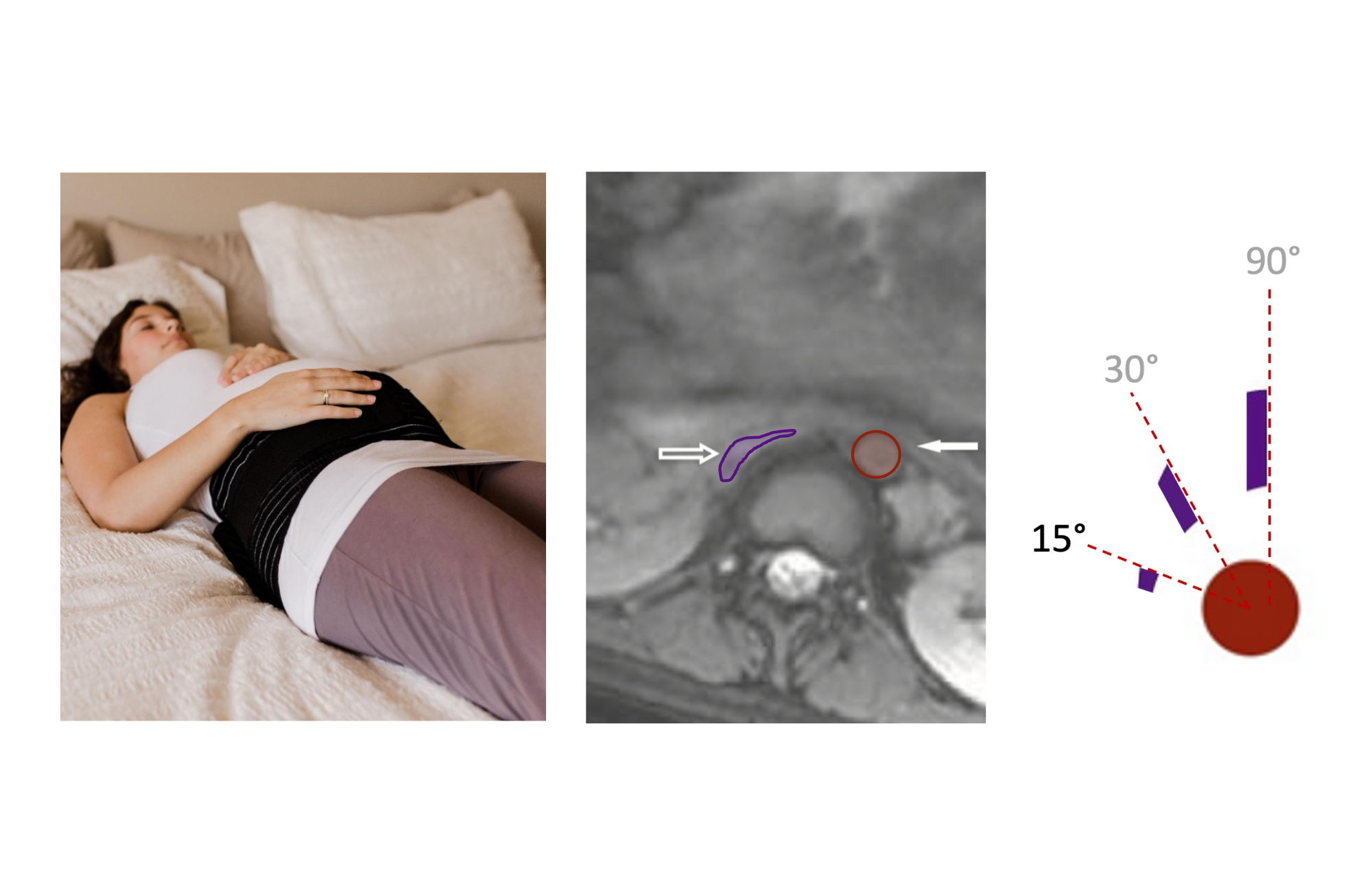
The PrenaBelt® arc-and-circle logo graphically represents the physiology of the effect of pelvic tilt in the third trimester of pregnancy on “aortocaval compression”. Aortocaval compression refers to the compression of two major blood vessels, the abdominal aorta and inferior vena cava, by the enlarging womb (uterus) in late pregnancy. These vessels are squeezed between the uterus and the bones of the spine. The abdominal aorta is an artery that carries oxygenated blood from the heart to the pelvis, uterus, and legs - this vessel is thick-walled and under high pressure and, therefore, can resist compression from the weight of the uterus. The inferior vena cava is a vein that carries deoxygenated blood from the legs, uterus, and pelvis back to the heart - this vessel is thin-walled and under low pressure and, therefore, is susceptible to compression from the weight of the uterus as the pregnancy advances and the uterus enlarges.

When a pregnant woman in the third trimester lies on her left side as seen in the photo below on the left, her uterus is no longer compressing her abdominal aorta or inferior vena cava. Let’s define this as 90° of pelvic tilt. The corresponding image on the right is an MRI showing a cross section of a 42 year-old pregnant woman lying on her left side at 39 weeks’ gestation. The image is taken at her L2-L3 disk level. The red circle from the PrenaBelt® arc-and-circle logo represents the cross-sectional area of the aorta and corresponds to the red overlay delineating the aorta on the MR image. The purple bar from the PrenaBelt® arc-and-circle logo represents the vertical height of the inferior vena cava and corresponds to the purple overlay delineating the inferior vena cava on the MR image.

As she rolls towards her back, her uterus begins to push on her abdominal aorta and inferior vena cava. The photo on the left demonstrates 30° of pelvic tilt. The corresponding MR image shows the cross section of a 39 weeks’ pregnant woman at 30° of pelvic tilt. The purple bar on the arc traces the vertical height of the inferior vena cava. Again, the red and purple overlays delineate the vessels on the MR image. The cross-sectional area of the abdominal aorta and inferior vena cava are essentially unchanged as the aortocaval compression at this angle of pelvic tilt is minimal.

As a she rolls further towards her back, her uterus begins to compress her abdominal aorta and inferior vena cava. The photo demonstrates 15° of pelvic tilt. The corresponding MR image shows the cross section of a 39 weeks’ gestation woman at 15° of pelvic tilt. The purple bar on the arc continues to trace the vertical height of the inferior vena cava, which is now rapidly decreasing due to compression from the uterus. Again, the red and purple overlays delineate the vessels on the MR image. The cross-sectional area of the abdominal aorta is still unchanged due to the muscular and thick wall of this high-pressure vessel. However, at this angle of pelvic tilt, the weight of the uterus resting directly on the relatively low-pressure inferior vena cava compresses it significantly.
Finally, when flat on the back in the third trimester of pregnancy, the uterus begins to compress her abdominal aorta and inferior vena cava. The photo demonstrates 0° of pelvic tilt. The corresponding MR image shows the cross section of a 39 weeks’ pregnant woman at 0° of pelvic tilt or flat on the back. The purple bar on the arc shows the vertical height of the inferior vena cava, which is now minimal. Again, the red and purple overlays delineate the vessels on the MR image. The cross-sectional area of the abdominal aorta remains unchanged. However, at 0° of pelvic tilt, the inferior vena cava is completely occluded by the weight of the overlying uterus.


Reference:
Higuchi H, et al. Effect of Lateral Tilt Angle on the Volume of the Abdominal Aorta and Inferior Vena Cava in Pregnant and Nonpregnant Women Determined by Magnetic Resonance Imaging. Anesthesiology, 2015.